Anatomy of the Hand & Wrist
The human hand and wrist are marvels of biomechanical engineering. These intricate structures enable us to interact with the world in remarkably sophisticated ways. Beneath the skin lies a complex network of bones, joints, muscles, tendons, ligaments, nerves, and blood vessels working together to carry out intricate tasks and movements. In this article, we explore the basic anatomy of the hand and wrist and their role in everyday movement.
Contents
The Anatomy of the Hand

The hand is an anatomically complex and intricate part of the human body. Here’s an overview of the key anatomical components of the hand:
Bones
The hand is made up of 27 bones grouped into three main sections: the carpus (wrist), the metacarpus (palm), and the phalanges (fingers and thumb). The wrist consists of eight small, irregularly shaped bones called carpal bones. Five metacarpal bones form the palm and extend from the wrist to the base of the fingers. Each finger has three phalanges (proximal, middle, and distal), except for the thumb, which has two.
Joints
Various joints contribute to the mobility and flexibility of the hand. They include the wrist joint (between the radius and carpal bones), the metacarpophalangeal joints (between the metacarpals and proximal phalanges), and the interphalangeal joints (between the phalanges). The metacarpophalangeal joints are responsible for flexion and extension movements. These joints allow the fingers and thumb to bend and straighten, enabling gripping, grasping, and pinching actions.
Muscles
The hand is equipped with a network of muscles responsible for the movement of the fingers and thumb, as well as overall hand function. These muscles control fine movements. The thenar muscles located at the base of the thumb help form thumb movements, including opposition and abduction. Extrinsic muscles located in the forearm and extending into the hand control major movements. The flexor digitorum superficialis muscle, for example, plays a key role in finger movement.
Tendons
Tendons connect the muscles to the bones and play a key role in creating the force needed for movement. The tendons in the hand work in conjunction with the muscles to facilitate intricate and precise movements. Fine motor activities, such as typing, writing, and picking up small objects, depend on the coordinated action of muscles and tendons.
Ligaments
Ligaments are strong fibrous bands that connect the bones and stabilize the joints. They provide support to the carpal bones and contribute to joint stability in the hand, limiting the range of motion of the joints to prevent hyperextension or hyperflexion. This restriction is essential for protecting the wrist and hand from injury.
Nerves
The hand is supplied by the median, ulnar, and radial nerves, which control sensory and motor functions. Sensory nerves transmit signals related to touch, temperature, and pain. Motor nerves control muscle movements in the hand and fingers.
Blood Vessels
The hand receives a blood supply from arteries and veins. This ensures oxygen and nutrient delivery to tissues and facilitates waste removal. Veins in the hand, including the superficial and deep veins, carry deoxygenated blood and waste products away from the tissues. This includes carbon dioxide, a byproduct of cellular metabolism, as well as other waste materials.
The Anatomy of the Wrist
The anatomy of the wrist is intricate, involving a complex interplay of bones, joints, ligaments, tendons, muscles, nerves, and blood vessels. The wrist connects the hand to the forearm and facilitates a wide range of movement. Below is an overview of the key anatomical components of the wrist.
Bones
The wrist is composed of eight carpal bones arranged in two rows. The carpal bones include the scaphoid, lunate, triquetrum, pisiform, trapezium, trapezoid, capitate, and hamate. These carpal bones provide structural support and flexibility to the bones of the forearm (radius and ulna) and the metacarpal bones of the hand.
Joints
The wrist joints connect the carpal bones to facilitate complex movements, such as bending and straightening. The radiocarpal joint is formed by the radius and the proximal row of carpal bones, while intercarpal joints are the joints between the individual carpal bones. The midcarpal joint is formed by the articulation of the proximal and distal rows of carpal bones.
Ligaments
The ligaments stabilize the wrist joints by connecting the carpal bones. Important ligaments include the radiocarpal, ulnocarpal, and intercarpal ligaments. For example, the ligaments in the wrist support the hand during weight-bearing activities, such as gripping and lifting objects. They distribute forces evenly across the carpal bones, reducing the risk of strain or injury.
Tendons
Tendons are fibrous tissues that connect the muscles to the bones of the wrist. The tendons of the wrist run along the dorsal (back) and volar (palm) aspects and facilitate movement of the wrist and hand. Tendons are important in fine motor control exercises, such as typing and writing.
Muscles
The muscles in the forearm control wrist movements by pulling on the tendons that cross the wrist joint. These muscles include flexors and extensors, each responsible for specific movements, such as bending and gripping.
Nerves
The nerves within the wrist include branches of the median, ulnar, and radial nerves. These nerves provide both sensory and motor functions to the hand and wrist. For example, the median nerve runs down the forearm and passes through the carpal tunnel, a narrow passageway in the wrist. It provides sensation to the palm side of the thumb, index finger, middle finger, and half of the ring finger.
Blood Vessels
Arteries and veins provide blood supply to the wrist. The radial and ulnar arteries contribute to the vascularization of the wrist and hand. In other words, they supply oxygenated blood to the hand and fingers.
When To Visit The Hand and Wrist Institute
Understanding the anatomy of the hand and wrist is essential for diagnosing and treating conditions or injuries that may affect this complex structure. Injuries to the wrist, such as sprains, fractures, and carpal tunnel syndrome, often involve damage to the bones, ligaments, tendons, or nerves within the wrist anatomy. Medical professionals, such as our Dallas hand surgeons, use their knowledge to assess and address a wide range of hand and wrist-related issues. Need an accurate diagnosis? Schedule an appointment with our orthopedic specialists today.
persons hand on black background by Nsey Benajah is licensed with Unsplash License

Dr. John Knight
Dr. Knight is a renowned hand, wrist and upper extremity surgeon with over 25 years of experience. Dr. Knight is a Board Certified Orthopedic Surgeon and Fellowship trained. Dr Knight has appeared on CNN, The Doctors TV, Good Morning America, The Wall Street Journal, The Washington Post, Forbes, The Huffington Post, Entrepreneur, Oxygen network and more.
Schedule Appointment

Meet the Doctor


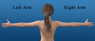
Where Does it Hurt?®
Not sure what service you need or what injury or syndrome you may have? Use our free, interactive tool to help you understand more about what you are experiencing. Start by clicking on the image below.
Real Patient Reviews

“I don’t often write reviews for Doctors offices..But this office is really exceptional in terms of service and my wrist is now great! Which is really the most important thing.”
Featured On