Kienböck’s Disease
Contents
- 1 What is Kienbock’s Disease?
- 2 What Causes Kienbock’s Disease?
- 3 What are the symptoms of Kienbock’s Disease?
- 4 How is Kienbock’s Disease diagnosed?
- 5 How is Kienbock’s Disease treated?
- 6 Surgical Options for Kienböck’s Disease
- 7 How can Dr. Knight help you with Kienböck’s Disease?
- 8 Animated Videos
- 9 Surgical Video
What is Kienbock’s Disease?
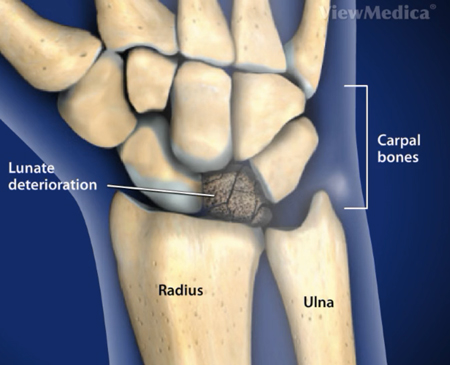
Kienböck’s Disease is a condition in which your lunate bone does not receive adequate blood supply. The lunate is one of eight small bones that make up the wrist joint. The lunate bone sits at the center of the wrist where it works with the radius and ulna in the forearm to support proper movement. Bones may seem fairly static, but they are living tissue. As such, your bones require a steady supply of blood to survive. Lacking the proper blood supply, the lunate bone will die and eventually collapse.

What Causes Kienbock’s Disease?
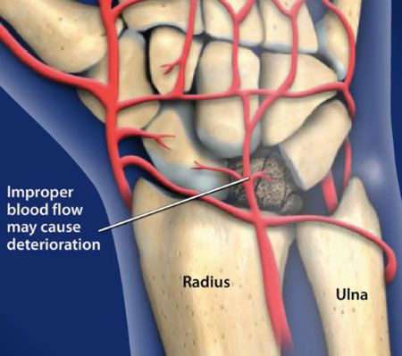
Kienböck’s Disease is caused by a lack of blood flow to the lunate, but it’s not always possible to determine exactly why this has occurred. Kienböck’s Disease is often precipitated by an injury that may damage the blood vessels in the wrist and compromise blood flow to the area. In these instances, patients often think they have a sprained wrist and don’t realize the severity of the problem until the pain worsens rather than goes away.
Some individuals are at a higher risk for Kienböck’s Disease because of the way their wrist is formed. In certain individuals, there are unique variations that can make it more difficult for the lunate bone to get enough blood, such as:
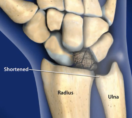
- An ulna bone that’s shorter than the radius bone.
- An irregularly shaped lunate bone.
- One blood vessel to the lunate rather than two.
If the bones in the wrist and forearm aren’t ideally shaped, your lunate may sustain more pressure from everyday activities than it otherwise would. This pressure can impact the blood flow and cause the lunate bone to collapse. If you have fewer blood vessels in your wrist, you’re also at an increased risk of suffering from poor blood flow to the lunate.
You may never know exactly what caused your Kienböck’s Disease, however, it’s important to promptly diagnose and treat the condition regardless of the cause.


What are the symptoms of Kienbock’s Disease?
Kienbock’s usually affects only one wrist and the primary symptoms include pain, stiffness, and reduced range of motion. The pain may vary in quality and duration and progressively worsen over time. Swelling and reduced grip strength may also occur.
Most patients go untreated for months or years. If left untreated the lunate bone may fragment or collapse. The abnormal lunate bone can wear away the ligament(s) making the wrist unstable and eventually resulting in Osteoarthritis.
As mentioned previously, Kienböck’s Disease can feel like a sprained wrist. Common symptoms of this condition in the early stages include:
- Wrist pain.
- Swelling of the wrist.
- Weakened grip.
In the first stage of the disease, an X-ray won’t reveal the problem. Symptoms at this time are usually mild, so you may not feel the need to seek medical attention initially. An MRI can detect the blood flow to your wrist, so this test will reveal the early signs of developing Kienböck’s Disease.
In the second stage of Kienböck’s Disease, the lunate bone begins to die and harden. You will experience worsening pain and may start to see swelling in the wrist. At this point, an X-ray will detect a problem as the dying bone appears different from the healthy living bone.
In stage three of Kienböck’s Disease, symptoms worsen noticeably. You may begin to experience:
- Wrist stiffness.
- Limited range of wrist motion.
- Tenderness on top of the hand around the middle of the wrist.
During the third stage, the dead bone breaks apart inside your wrist. This causes the surrounding bones to shift inward. At this point, your wrist may no longer have the strength or support to function properly.
The lunate bone collapses completely in the fourth stage of Kienböck’s Disease. The surrounding bones and connective tissues may sustain damage as a result, which will cause all of the previously mentioned symptoms to become more severe. Progression from stage one to stage four can take anywhere from a few months to a few years.
How is Kienbock’s Disease diagnosed?
A detailed medical history and review of symptoms is the first step towards an accurate diagnosis. Kienbock’s is more difficult to diagnose in the early stages since the symptoms can mimic those of a sprained wrist. Imaging studies like MRIs and X-rays are extremely useful in diagnosis and staging of this disease.
How is Kienbock’s Disease treated?
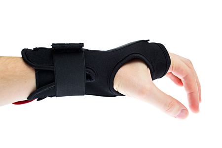
In the early stages of Kienböck’s Disease, you can address pain and swelling with anti-inflammatory medication like ibuprofen or aspirin. As the condition worsens, you may need to modify your activities or immobilize the wrist. Your doctor may recommend splinting the wrist as needed. Alternatively, you can have the wrist immobilized in a cast for two to three weeks to relieve pressure on the lunate bone. In most cases, you will eventually need to pursue surgical treatment for Kienböck’s Disease.
Non-surgical
If caught early enough surgery can be avoided. Anti-Inflammatory medications such as ibuprofen can relieve pain and swelling. The wrist may need to be immobilized in a cast or splint for several weeks or months while healing occurs. If symptoms do not improve, surgical intervention may be required.

Surgical Options for Kienböck’s Disease
There are five primary options for Kienböck’s Disease surgery. Your surgeon will choose the best approach based on your symptoms, activity level, and personal health. Options include the following.
Revascularization
In the early stages of Kienböck’s Disease, you can pursue revascularization surgery to restore blood flow to the lunate bone. This may cure Kienböck’s Disease and prevent further damage to the wrist. Revascularization is done by taking a piece of bone from another part of the forearm or wrist that still has blood flow and grafting it onto the lunate bone. The wrist is usually immobilized during the healing process using a metal plate connected to pins that are inserted into the bone.
Intercarpal Fusion
Intercarpal fusion fuses the lunate to the carpal bone. This can be combined with revascularization to restore blood flow.
Joint Leveling
If your forearm bones are of differing lengths, your surgeon can lengthen or shorten bones. This relieves pressure on the lunate.
Lunate Excision
Excision is the removal of the lunate bone. The surgeon usually performs a partial wrist fusion at same time.
Proximal Row Carpectomy
A proximal row carpectomy removes four of the eight wrist bones. This procedure is used in the final stage of Kienböck’s Disease after the lunate bone has collapsed. Removing the lunate and surrounding bones can alleviate pain while maintaining partial range of motion in the wrist.
Early Stage:


Late Stage:


How can Dr. Knight help you with Kienböck’s Disease?
When it comes to rare and complex wrist issues like Kienböck’s, it is important to find a surgeon who is well-versed in their treatment and who has seen many cases. Dr. Knight is one such surgeon, and he will bring his expertise to bear in your case, which will, no doubt, lead to the restoration of function in your hand and wrist.
We’re looking forward to helping you live a more pain free life. To schedule an appointment with Dr. Knight, contact us online or give us a call at (855) 558-4263. With offices in Dallas, TX and Southlake, TX, we are able to serve the greater Dallas Fort-Worth area and surrounding communities, including Irvine, Arlington, Lewisville, Carrolton, and more.
Animated Videos
Surgical Video
Note: The following video contains graphic images.
Disclaimer
HandAndWristInstitute.com does not offer medical advice. The information presented here is offered for informational purposes only. Read Disclaimer


