Cartilage and Ligament Tears of the Wrist
Contents
- 1 What are Cartilage & Ligament Tears of the Wrist?
- 2 What causes Cartilage & Ligament Tears of the Wrist?
- 3 What are the symptoms of Cartilage & Ligament Tears of the Wrist?
- 4 How are Cartilage & Ligament Tears of the Wrist diagnosed?
- 5 How are Cartilage & Ligament Tears of the Wrist treated?
- 6 How can Dr. Knight help you with cartilage and ligament tears?
- 7 Testimonials
- 8 Frequently Asked Questions:
- 9 Surgical Videos
What are Cartilage & Ligament Tears of the Wrist?
The wrist is made up of 8 small bones (carpals) arranged in two rows of four bones each. The proximal row of carpals articulates directly with the bones in the forearm called the radius and ulna. The distal row connects to the long bones (metacarpals) in the palm of the hand. Linked together like a chain, the two rows of carpal bones allow the hand to move up (dorsiflexion), down (palmarflexion) and from side to side (radial and ulnar deviation).
Each carpal forms a joint with the bone next to it. Articular cartilage covers the ends of each bone at each joint. Cartilage is the tough slippery white substance that allows bones to glide past one another without damage. Ligaments are strong cord-like structures that connect bone to bone. The ligaments of the wrist not only attach the carpal bones to one another, but they connect the carpals to the radius and ulna, as well as, to the metacarpal bones.
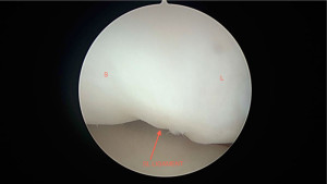
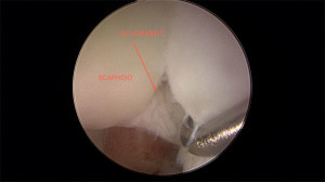
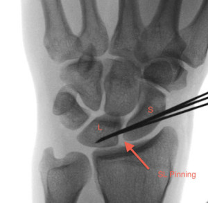
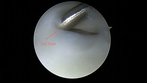
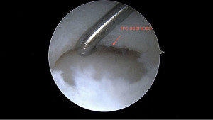
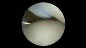
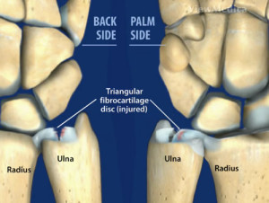
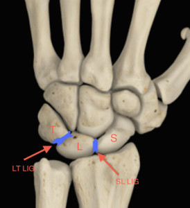
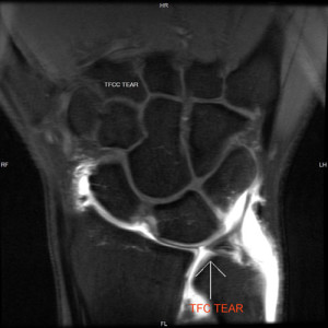
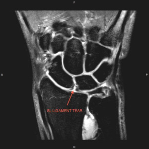
The cartilage and ligaments that unite the proximal wrist are the most prone to injury. The triquetrum (T), lunate (L), and scaphoid (S) are the carpal bones of the proximal row. The ligaments connecting these bones are the luno-triquetral ligament (LT) and the scapho-lunate ligament (SL). The triangular fibrocartilage complex (TFCC) is made up of the cartilage and ligaments that suspend the proximal carpals in place against the ulna and radius. The triangular fibrocartilage (TFC) is the cartilage that articulates primarily between the ulna and triquestrum, and the edges of the radius and lunate bones (See Picture). The TFCC provides stability to the wrist and is a focal point for force.
What causes Cartilage & Ligament Tears of the Wrist?
Acute trauma or repetitive use over time can lead to tears in the cartilage and/or ligaments. The most common cause of injury to the cartilage and ligaments of the wrist is a hard fall on an outstretched arm with the palm side down or from excessive torque at the wrist. If the ulnar side of the wrist is hyperextended the TFCC could tear or rupture. Gymnasts, tennis players, golfers and other athletes are at an increased risk for this type of injury. Additionally, any type of wrist injury can alter the stability of the joint leading to increased forces acting on the ligaments and cartilage. Over time the damage from these forces is cumulative and the joint can no longer compensate resulting in pain and diminished range of motion. Degenerative tears can also occur secondary to inflammatory disorders and some congenital variances.
What are the symptoms of Cartilage & Ligament Tears of the Wrist?
The most common symptoms are pain and swelling of the wrist. The pain may increase with activity or a specific movement. In acute injury the site may become bruised or discolored. Patients report weakness, reduced range of motion and occasionally a clicking or snapping sound. Overtime this injury may lead to arthritis of the wrist with increased stiffness and pain.
How are Cartilage & Ligament Tears of the Wrist diagnosed?
After a complete history including occupational hazards, review of symptoms, and prior trauma a complete a physical examination will be performed. The doctor will assess for grip weakness, edema, alignment, joint stability, excessive movement, and range of motion. X-rays show the presence of bone fracture and the alignment of the bony structures in the wrist. Arthrography is used along with an MRI with an injectable dye to determine if tears are present. If the dye leaks into the joint the test is considered positive. MRI can visualize the ligaments and other soft tissues to detect damage or rupture.
How are Cartilage & Ligament Tears of the Wrist treated?
Non-surgical
If the wrist is stable, tears to both ligaments and cartilage can be treated by immobilizing (splinting) the wrist for 4 – 6 weeks. NSAIDs (non-steroidal anti-inflammatory medications) such as ibuprofen may be taken to relieve pain and inflammation. Steroid injections and physical therapy may also be prescribed.
Surgical
If the injury is severe or does not respond to conservative treatment then surgery is indicated. Wrist Arthroscopy can often be used to diagnose and repair both ligament and cartilage ruptures.
Ligament Tears
The ligaments of the wrist begin to heal, tighten, and scar after injury. The accumulation of scar tissue may interfere with the re-alignment of bone and change the scope of the surgical intervention. Rupture to the ligaments of the wrist can be repaired by pinning, reconstruction, or fusion depending on the severity and amount of time that has lapsed since injury.
If surgery is performed within three months of injury pinning may be the best option. Pinning refers to metal pins that hold the bones in place while the ligaments heal. This can be accomplished percutaneously (through the skin) or thru a tiny incision, to first repair the ligament. The pins are then removed in approximately 8 weeks.
In cases where arthroscopic surgery is unsuccessful or the ligament injury is discovered long after the initial injury, arthrodesis (bone fusion) may be indicated. Bone fusion is achieved by removing the cartilage between the bones causing them to grow together as they would in the case of a fracture. This reduces pain from friction in the joint and stabilizes the bones. Unfortunately fusion can result in a significant loss of range of motion.
Triangular Fibrocartilage Complex Tears
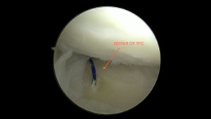
Most repairs to the triangular fibrocartilage can be performed arthroscopically. Depending on the severity and location of the tear the procedure may require debridement (cleaning) or a repair. Post-operatively a splint or cast may be worn for a week if debrided or for 6 weeks if repaired. Dr. Knight will explain the specifics of your individual surgery.
How can Dr. Knight help you with cartilage and ligament tears?
Dr. Knight is one of the leading specialists in his field in the treatment of wrist injuries and disorders. He has significant experience in making the difficult diagnosis of a cartilage and ligament tear and has a high success rate with minimally invasive stitchless advanced wrist arthroscopy in returning the elite athlete to their sport.
Come visit Dr. Knight, one of the most accomplished hand specialists in Dallas, Texas. See him at the Southlake office or Dallas hand and wrist center.
Testimonials
Cartilage Tears of the Wrist Testimonial
A few years back I severely injured my wrist skiing. The injury resulted in frequent dislocations and a lot discomfort. The instability was affecting my everyday life and especially my skiing. I eventually decided it was time to see a doctor. I extensively searched the Internet looking for the best doctor and I found Dr. Knight. From our first introduction I instantly felt very comfortable and confident I had made the right decision in choosing him.
The initial diagnosis was not good. The MRI showed multiple torn ligaments in my right wrist. Dr. Knight was very upfront and informed me that since my injury had occurred a few years back, there was a very good possibility that the ligaments may be damaged beyond repair, and the success rate of reconstructive surgery dropped significantly. He in fact could not recall anyone waiting as long as I did to go through with reconstructive surgery on an injury as severe as mine. With skiing and surfing as one of my main priorities in life I decided that the best option was to take the chance and go forward with the procedure.
I am very happy with the decision I made. Dr. Knight was able to repair my wrist and 6 ½ months later I was able to surf again and I was back on my skis and able to push it on the mountain without any limitations.
David Soroka
Frequently Asked Questions:
What exercises can I do to help with ligament tears?
It is important not to overextend or overwork the ligaments of the wrist during recovery, but under the guidance of an occupational therapist, there are many exercises that can help your injuries heal faster and with less loss of function. Flexion and extension exercises are where the wrist is bent forward and then backwards, or towards the ground, followed by towards the sky. These will help make sure the flexor and extensor tendons keep their flexibility. Side-to-side stretches will work the same tendons, abut on the opposite orientation, to increase range of motion.
How long does wrist ligament surgery take to heal?
Recovery times vary based on severity and extent of the injury, but after a surgery to repair a torn ligament in the wrist, you will, conservatively, need at least six weeks for the tissues to fully heal themselves. The wrist is a very delicate part of the body, and so it is important that the recovery is carefully monitored, to avoid complications that might lessen use or decrease range of motion.
Is there a special brace for people with TFCC tear?
As TFCC tears do not generally require surgery, splinting or bracing is an important part of the recovery process, and there is a particular kind of brace that is used in these cases, that has a hole in it where the ulna’s protruding head can rest, while the rest of the brace holds the wrist bones in proper alignment for healing.
Surgical Videos
Note: The following videos contain graphic images.
Disclaimer
HandAndWristInstitute.com does not offer medical advice. The information presented here is offered for informational purposes only. Read Disclaimer

