Hand Fracture Surgery
Transcript:
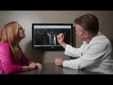
Let’s take a look at hand fracture surgery the hand is very susceptible to injuries from direct blows to crush injuries to torquing injuries to falling on an outstretched hand let’s look at some of the common one though this is a martial artist proper punching is key but frequently martial artists and boxers and those of us that uh maybe have a little anger management issue and go out and strike a wall occasionally are susceptible to improper technique and can get uh fractures of the hand also Crush injuries let’s look at the uh Carpenter here just H hammering nail after nail can slip causing a crush entry particularly to the tip of the finger now let’s look at the anatomy that’s involved with finger fractures first we need to look at the bones of the hand where are they well the the fingers are made up of fanges and there’s three fanges distal middle and proximal failinks of all the fingers the thumb has just two the distal and the proximal fail lengths and then there’re the metacarpal bones the metacarpal bones are the bones that bridge between the knuckles and the wrist you can see there’s five metacarpal typically labeled one through five starting with the thumb index middle ring and little fingers so let’s take a look actually now at the actual fracture patterns there the metacarpal fractures they can be transverse in orientation just straight across they can be oblique or more of a spiral fracture where they kind of wrap around the bone or they can be again a short oblique and the oblique fractures typically are inherently unstable because the forces across the fracture typically displace this so they’re usually highly unstable now let’s take a look at actual uh metacarpal it’s called a boxer’s fracture which is a metacarpal neck fracture this again is from improper punching striking a wall or another human’s head and what happens is the the knuckle when it breaks it’s out right near the joint and again fractures displace because the muscles and tendons pull them in a Direction they shouldn’t be going so the typical tendency is for that bone or the head of the metacarpal to go in a palm word direction and these typically we can in most cases we accept a lot of angulation because fixing these fractures would typically cause more stiffness than not. So uh we’ll accept up to 70° of angulation with these but in some as you’ll see in a minute you actually have to put some pins in. Now take a look now at typical fracture patterns of the me of the in the fingers. On the top left is called a tuft fracture the distal failinks and this again occurs usually from a blow from a hammer or a crush injury and usually can rip apart the nail and the skin and the bone actually can protrude through the skin and is needs to be repaired right away many times in the emergency room by simply sewing back the the nail bed and placing in a splint. Sometimes these do need internal fixation as we’ll see in a minute. On the bottom left this is the proxim failen fracture called No Blake fracture because it’s on a bevel and again from the deforming forces from the tendons typically angulate or displace and need to be fixed. Up here the middle failinks again uh this is a transverse fracture but the tendons that extend and bend the and flex the finger typically pull this in different directions again leading to an unacceptable alignment. And finally an oblique fracture of the proximal failinks out towards the head you can see it’s on the on the angle and again is displacing. So these fracture the fractures we’re talking about today are ones that typically need to have uh surgical repair or internal fixation. now let’s take a look at at the clinically what we look at obviously if the bone comes through the skin we see that and we see deformity if the uh if the finger is crooked but one Telltale sign that’s very important on fractures is a look at the finger alignment. The finger should bend down towards the base of the thumb here all in unison. The little finger a little bit under the ring finger but basically are all on Unison. When you break a bone that the uh the metacarpal or the fanges the bone can twist uh and it can lead to a a deform what we call a rotational deformity of the finger. This is 101 fracture treatment and it must that must be properly aligned so it’s one of the first things we’ll do is is even though it’s broken we’ll have you bend a little bit so we can gauge if there’s rotational Mal alignment so take a look at the little finger here you can see it underlapping or overlapping the adjacent finger so this can be very debilita a with use of the hand. Let’s look at this in an actual actual individual. This gentleman was a musician. He had broken his finger didn’t get medical treatment right away for the first couple months. It healed in this crooked position but look at the significant deformity here all from a break right here. So he had to go back in his case rebre the bone reposition put a plate and screws in to correct that because as a musician he had a lot of problems with this deformity. Now let’s take a look now at some before and afters but before we do that just to you some typical fixation. The we we’ll talk about two predominant fixation. one is just percutaneous putting a wire through the skin to uh reduce to hold the fracture in place which is done under a special fluo or uh IM real-time Imaging that we use in surgery to see where the pen is going and to make sure the bone is properly aligned. But in some fractur those ones where where they’re typically long oblique fractures that are very very unstable we actually have to cut open the patient and put some screws in place. The problem with that is it the more cutting you do the more the scarring and overall rehab time and potential for other surgeries to release adhesions. So now look at some before and afters. This next uh is a cap scan of an individual who struck a a um a heavy bag in boxing so hard that he dislocated the third fourth and fifth metacarpal. This is a CAT scan showing what we call fracture dislocation. So the base of the third metacarpal here where the arrow is you can see the fracture goes into the joint but the whole joint is dislocating. So all three of those joints uh at the base of the middle ring and little fingers were dislocated or shifting out of place. So we had to go in and put those back in place. So let’s look at the after effect look at all the pins but here are the three joints down here that were all popped out of place and we went in and and place these back in place with with an incision because we had to open open and put these back in perfectly and then we secured these with multiple pens about 5 Weeks Later 5 to 6 weeks later typical healing time little minor surgery take the pens out and then started rehab. Now let’s take a look now at a thumb metacarpal fracture. This is a fracture from usually from a jamming or torquing injury to the thumb. And it you can see the base of the metacarpal here and because of all the the muscles that are bending the thumb and pulling the thumb in different directions this bone tends to angulate. So in this individual we went in put the bone back in place place without having to cut open the patient and putting a couple pens across. So let’s look at the after uh x-ray here you can see good alignment. It now it’s straight not crooked two pens across again for about five weeks and those come out and start therapy. Now let’s take a look at at the we talked to you earlier about that about that boxer’s fracture. Typically we accept a lot of angulation. But one time one time that we do fix these if the bone is completely separated. Look at the look at the shifting of the head of the bone down here here in relation to the shaft of the bone it’s completely knocked off as as we say. So in this individual we went in put it back in place again without having to cut open and then shot a couple pin across as you can see in the next x-ray right here. Uh you can see two pens going across the fifth metacarpal head into the fourth metacarpal head just anchoring that back in place. Then the pins come out and again more rehab. So let’s take a look now at another fracture. This is an individual that was in a motor vehicle accident and he sustain fractures of the third fourth and then over at the base of the little finger towards the head another fracture but that was non-displaced. It’s really the the third and and the third and fourth metacarpal here had shortened and and the patient had rotation of the fingers. So again simple in this case we had to go in and put some screws in and do it through a small incision on the back of the hand and we move the tenons out of the way. Then we go and put two screws in. Let’s take a look at the next x-ray you can see two screws here and two screws here. We lag those in so as we tighten them up that bone compresses. And you can see you know much better alignment now the the the preservation of the length of the bone and clinically he had no rotation of his fingers like we showed in that individual earlier. Now the next fracture we’re going to look at is a fifth metacarpal fracture again just to show you more of the same really but to show you different fracture patterns. This is long oblique fracture with the uh with the pulling of the tendons tends to shift the finger in this direction. So in this individual again went in cut him open put two screws in. Let’s take a look so we put the screws in from two different angles going in two different directions to compress that fracture with the best um from the best position possible. Now another fracture we’re going to look at is this is in a professional snowboarder uh who uh qualified for the Olympics. Before doing so though she had a horrific accident where she fell uh sustaining a fracture of the index or the second metacarpal. This is a CAT scan here. And you can accept very little angulation of the fractures as we go into the index and middle fingers as opposed to the the ring and Little Finger metacarpal. So this is unacceptable angulation several pieces. And we went in and put a plate in screws as you can see on the next x-ray right along the back side or dorsal side of the metacarpo and she did great return to professional uh snowboarding. Now the next uh fracture we’re going to look at is a school teacher that had her little finger pulled in an outward Direction looks very painful and it was. Look at the displacement of the F of the fracture right here and she came in with her finger literally pointing almost 90 degre to the outside. So we in this individual these are great for just putting in P percutaneous pin without having to do any cutting. So we take her to surgery put her to sleep push that back in place under that special fluo Imaging device and then shoot two pens across as you can see here crisscrossing. These are interoperative X-rays. You can see crisscross the the normal alignment of the finger and she did very well from this. Now let’s look further into into the finger fractures. Look at look at this great image right here showing this significant fracture of the index uh proximal Phil length and the fracture extends into the joint but this was not separated so we were actually lucky and not having joint involvement. Went in cut her open on the back of of the proximal fail lengths split the tendon and then put two p uh screw. Let’s look at the after effect here you can see two screws right here compressing the fracture with anatomic alignment and these are low these are low profile um 1.5 millim titanium screws so they’re designed to stay in. Occasionally if one loosens you’ll take it out uh but usually that’s all that’s involved. Okay so let’s take a look now at the this individual had a middle failen fracture from a jamming injury. Initially she had a non-displaced fracture. So you ask your hands why you seeing me so often well one reason is because if the fra that hairline fracture or non-displaced fracture starts to shift we want to catch it before it heals. So typically you’ll come in every two weeks we’ll get x-rays so at about three to four weeks she actually started to shift and the bone the finger started deviating and it was deviating as you can see the ring finger towards the little finger over here. So we went in surgically put her to sleep put it back in place no cutting actually and just were able to put three pins in. Let’s take a look at the uh pins here. We put one across the uh the base of the or the tip of the bone here just to stabilize The Joint because that crack actually extended into the joint so we didn’t want to displace that and cause more of a problem. Then we crisscrossed two pin here holding this in normal alignment. Now pin came out about five weeks later. Let’s take a look at the aftermath here you can see right here uh where the previous pens were which you’ll heal in but this is right after the pin were removed and you can see normal alignment of the digit. Okay so the uh one more fracture I want to look at this is a base of the proximal fail length fracture. This shows a good um good uh pict picture of the fracture going into the knuckle or into the articular surface. This this is slightly separated but in a very important joint like the metacarpal fangel joint or the knuckle. You need this as perfect as possible and this individual had some separation here but it also had a rotational deformity. So we knew we had there were many reasons to look at doing surgery this the main one was a rotational abnormality and then and then also there was some joint involvement. So we went in made an incision split split the tendon and then in this case because we lot of little pieces it was not a minable to putting those um those screws that could stay in. So in that this case we went in and put some wires. Let’s take a look we went in this patient also had a rotational abnormality so many reasons to fix this but went in surgically put open the joint up put everything back in position put several pins in because they were so many small pieces the screws uh were not adequate for this. So we felt based on the fracture configuration we put pins in and these would stay in uh in this individual about 6 weeks and then the come out and then he would need you know quite a bit of Rehabilitation after that. So after these types of uh finger fra or hand fractures usually there’s a period of immobilization and a you usually a removable brace for about four to six weeks. Then a minor trip to surgery if we put in pens to take those out. It’s usually like a f minute outpatient procedure with a Lo little local anesthetic little IV sedation for your comfort. Once these pins come out you’re look or or the fracture is healed 4 to 6 weeks later you’re looking at up to 2 to 3 months of of of pretty intense rehab to get the movement back. But the fingers are so important to the function of the hand you got to get there. For more on this condition and many other conditions please check out our website.
Schedule Appointment

Meet the Doctor

Where Does it Hurt?®
Not sure what service you need or what injury or syndrome you may have? Use our free, interactive tool to help you understand more about what you are experiencing. Start by clicking on the image below.
Real Patient Reviews

“I don’t often write reviews for Doctors offices..But this office is really exceptional in terms of service and my wrist is now great! Which is really the most important thing.”
Featured On