Cubital Tunnel Surgery
Transcript:
Let’s talk about cubital tunnel surgery. Cubital tunnel syndrome is compression of the ulnar nerve at the elbow. It’s the second most common area of nerve compression, secondary only to carpal tunnel syndrome. There are many daily activities that can precipitate cubital tunnel syndrome. Let’s look at one right here. This is a lady talking on the phone, bending her elbow. This repetitive elbow flexion or bending for prolonged periods of time can cause increased ulnar nerve. Also as this this gentleman right here is doing, he’s resting his elbow on a hard tabletop. Not a good thing. We all have poor posture and tend to lead lean to one side or the other just as he is doing and this can also cause problems. Also sitting at the keyboard all day long typing and moving the hand and fingers and the wrist repetitively can lead to increased tension or inflammation around the ulnar nerve at the elbow causing symptoms. Let’s take a look at the anatomy though to get a better appreciation of what’s happening here. The ulnar nerve is this yellow structure coming down the inside of the arm right along here. Goes right behind the medial epicondyle at the end of the humerus bone or what we lay people refer to as the funny bone. It’s in this area behind the funny bone or epicondyle here and can get entrapped. And then what this nerve does, the function is it gives sensation to the little and ring fingers. But more importantly, it gives us all the motor fine dexterity or fine muscle movement within the hand. Most of the muscles are supplied by the ulnar nerve that gives us the ability to pick up small objects, coins, button clothes, anything force, any pinching of keys and things like that. This nerve gives us the motor, the muscle function, or impulse for that muscle function. So let’s take a look at now the area of compression. The nerve comes down behind on the inside of the arm, goes behind the funny bone. This is the epicondyle. Muscles that flex the fingers or bend the wrist all come from this area. So it’s beneath behind this bone where the nerve can get with flexion of the elbow gets pulled up behind that area leading to compression. Also the nerve then comes between two muscle bellies right here with this band of tissue.
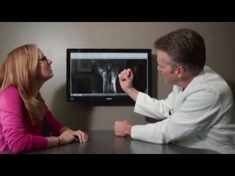
That it’s in this area where that tunnel can become compressed and cause symptoms. So let’s take a look at the actual symptoms that are happening. Classically numbness and tingling with cubital tunnel syndrome affects a little finger and half of the ring finger. Now pain also can occur from along the inside of the elbow at the point of compression and with this rectangular box is an area where where patients can feel symptoms of pain up and down the forearm. In severe cases, the circle areas are those fine little muscles that give us dexterity and these areas in severe cases you can get weakness or loss of pinch or grasp or these fine motor movements with almost with paralysis in really severe cases. So let’s take a look now at the what what we need to do as a hand specialist to diagnose the problem. First and foremost, this is a clinical, a detailed clinical examination is what leads to the diagnosis. That’s the most important thing. But then we’ll get electrodiagnostic testing or what’s referred to as an nerve conduction velocity test. And what the neurologist does that we send these patients to is they put an electrode in the hand and the stimulator here and they’ll stimulate the nerves down the forearm, across the elbow and across the wrist. If there’s any delay such as in cubital tunnel syndrome between above the elbow to just below the elbow, that’s indicative that there is a lack of impulse or a lack of circulation to the nerve causing cubital tunnel syndrome. It’s important to know though in 20% of patients a normal electrodiagnostic testing may be present in a patient that has clinical findings of cubital tunnel syndrome. And on the other side of the coin, in 20% of patients that the nerve test shows cubital tunnel syndrome, the patient may not have any symptoms. So it’s very important to first and foremost as a hand specialist to examine the patient. That’s how we make the diagnosis. And then we can look at the nerve test as an adjunct to determining just how severe the problem is. Okay, let’s take a look now at at the treatment for this. Conservative treatment at the top here. Resting the arm in overused syndromes or overused conditions, putting the arm, the elbow in this case at rest as well as.
The wrist and hand. Needs anti-inflammatory medication to reduce inflammation within that tight space. Ergonomic modifications. We look at again as we said resting the elbow on hard objects, so we want to get that elbow off of the arm the the um the armrest of the chair. Have it just supporting the forearm. A comfortable position at the keyboard. And then modifying the amount of tapping, amount of activity, amount of repetitive movement of the elbow, wrist and fingers. We want to reduce that by ergonomic modifications. And finally the most important bracing technique that can help this is called a pillow pillow brace. In the old days, people used to take a brace or a pillow, wrap it around the arm to keep the elbow from bending at night. The nighttime is a time when we rest. We want to rest the elbow. We tend to sleep in a fetal position bending the elbow which puts undue stress across the nerve. So we want to get a pillow brace. It’s spelled P I L hyphen O brace. Just Google it. You’ll see many places you can order this. And what this does is it holds the elbow in a in extended resting position at night. So if conservative treatment fails, then we have to look at surgical intervention. There’s some three techniques that are highly successful for surgical intervention. But let’s take a look at what again the anatomy. The ulnar nerve coming down behind the epicondyle. And the first treatment with surgery is to go in, as you’ll see from this next image, looking at it from the side view. We want to go in here and we want to open up the cubital tunnel. Open up those sources of compression. Then once we open it up, we’re going to do one of three procedures. Typically these are the standard procedures. The first two we’re going to talk about are called transposition procedures. As you’ll see in this in this case, the nerve is decompressed and then the nerve is going to be moved over the epicondyle. There’s two types. One is putting the nerve below the muscle which is called a submuscular transposition. And the other is putting it above the muscle and that’s called a subcutaneous transposition. Both of these move the nerve from its original bed and put it on, take it with the blood vessels and put it on top either top of the muscle or below the muscle. Now, below the muscle or submuscular.
Submuscular transposition has been advocated for elite overhead athletes such as pitchers in baseball or professional quarterbacks because the velocity they throw the ball or football with is so significant that it puts a lot of stress across that area. So you want to put the nerve in that case below the muscle to really try to protect it. So again, we’re going to move the nerve forward and as you’ll see here, we you the nerve is now in a different course. So the nerve comes down, it was back here, now it’s been flipped up and it’s going either above the muscle or beneath the muscle diagonally across the elbow. So the this next slide will actually show a nice animated image here. The nerve is being decompressed. This yellow structure, muscles being elevated. The nerve is put down through that. So those are the two transposition procedures. But there’s one third procedure that’s very popular and that’s decompressing the nerve as we said right behind the funny bone. And then we’re actually going to remove half of that funny bone or the epicondyle. The nerve and then if we just did that, the nerve could flip back and forth when the elbow bends or or stands and so we don’t want that to happen. That can cause problems with the nerve. So we take off a little bit of the bone just to narrow, just to remove part of the hill that nerve has to climb over. And but we we it’s very important to not take off too much bone or you can disturb the all important ulnar collateral ligament. So after this, we’re going to take a look at now in an actual patient. This is towards the hand. Up up that way is towards the head. So here comes the ulnar nerve as you can see denoted by this arrow here. Little blood vessels with it. Nice picture of this. And this is the epicondyle. Muscles of the FCU muscle. And it’s between these two thick bands that nerve can become compressed such as this case. So we’re going to show this. We’re going to ultimately open up this tunnel completely. And then we have we can do one of those three procedures. Either take this nerve, cut a trough for the muscle, put it beneath the muscle, simply just put it on top of the muscle and anchor it with some of the fat right here. Or we can elevate the muscle from this bony prominence or the ep.
Epicondyle. And then the nerve actually just stays in its normal position. So after the surgery, we’ll place you in a in a long arm splint from the fingertips all the way up above the elbow but we leave the room for the fingers to move. After the with the splint on for the epicondylectomy procedure, you’re usually in a brace for about 10 days. Then the splint comes off and you start therapy. In the transposition procedures, usually you’re immobilized for three weeks. Overall, the total recovery time following this procedure is about 2 to 3 months. The overall success rate is about 90%. For more on this condition and many other conditions, please check out our website.
Schedule Appointment

Meet the Doctor

Where Does it Hurt?®
Not sure what service you need or what injury or syndrome you may have? Use our free, interactive tool to help you understand more about what you are experiencing. Start by clicking on the image below.
Real Patient Reviews

“I don’t often write reviews for Doctors offices..But this office is really exceptional in terms of service and my wrist is now great! Which is really the most important thing.”
Featured On