Colles Fracture: Treatment & Splinting
What is a Colles Fracture?
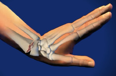
A true Colles Fracture is a complete fracture of the radius bone of the forearm close to the wrist resulting in an upward (posterior) displacement of the radius and obvious deformity. It is commonly called a “broken wrist” although the distal radius is the location of the fracture, not the carpal bones of the wrist.
Fractures of the distal radius are extremely common and historically several methods of classification have been proposed. At present time in the United States, and for the purposes of this article, we will refer to all distal radius fractures as Colles Fractures. Smith fractures, Chauffer’s fractures, and Barton’s fractures- other types of distal radius fractures- are also included under the umbrella of distal radius fractures.
Distal radius fractures are further classified based on certain characteristics. A fracture of the distal radius can be intra- or extra-articular, depending on whether the fracture extends into the wrist joint. These intra- or extra-articular fractures can either be displaced or alignment can be maintained. When the distal radius is broken into many small pieces of bone as in a crush injury, it is deemed a comminuted fracture. Distal radius fractures can also pierce the skin resulting in an open fracture.
What are the causes of a Colles Fracture?
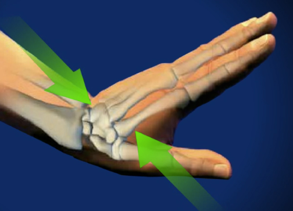
Although Colles Fractures are a heterogeneous group of injuries, the etiology is usually the same: a fall on an outstretched arm. The position of the hand is important in determining the type of fracture that results. For example, in a true Colles fracture, the fall occurs in the forward direction with the wrist extended to break the fall, and in the rarer Smith’s fracture, the fall occurs onto a flexed wrist.
Colles fractures can also be caused by direct trauma.
There is a bimodal peak of incidence of Colles fracture. High energy falls, such as from a motorcycle, occur in the 18-25 age group, and low intensity falls, such as from tripping and falling occur in senior citizens with brittle bones.

What are the symptoms of a Colles Fracture?
Pain, swelling, bruising, and deformity are common symptoms of a Colles Fracture, depending on the type of fracture and its characteristics.
How is a Colles Fracture diagnosed?
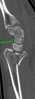
A careful history including the mechanism of injury establishes suspicion for a Colles fracture. Physical examination assesses the soft tissues surrounding the joint and determines if the surrounding nerves and blood vessels have been injured. X rays and/or CT scan will confirm the presence and classification of the fracture.
How is a Colles Fracture treated?
Treatment of Colles fractures depend on the type of Colles fracture present, the age and activity level of the patient, the surgeon’s preference in treatment method, and the patient’ s desires regarding immobilization and return to activity. As Colles fractures are so common, many methods of treatment have been developed to stabilize the fractures and allow the bone to heal. The goal is to return the wrist to its prior level of functioning.
Non-surgically
If the fracture is not displaced and pain is minimal, a simple splint may be used to immobilize the area. This method is most frequently employed in elderly, low activity level patients. In young, active patients a plaster cast is usually placed to ensure immobilization and proper healing.
If the fracture is minimally displaced, the surgeon may manually move the bones back in place in a procedure called closed reduction. This is followed by a plaster cast for 6 weeks during which time x rays are taken periodically to ensure the bone remains aligned.
Surgically
Surgery is indicated for displaced fractures or fractures that are unstable and fail closed reduction. There are several surgical techniques available.
In closed reduction and percutaneous pinning, the pins are inserted into the fracture, manipulated to realign the bones, and then used to stabilize the fracture. The pins are removed after the fracture has healed.
In external fixation, a device known as an external fixator is used to pin the bones in place and hold them steady. The pins are attached to a device worn outside of the wrist. Numerous models of external fixator have been developed for distal radius fractures over the years.
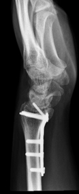
Plates are often employed to stabilize the Colles Fracture with excellent results. The plates may be placed on the front or volar side of the wrist or the back or dorsal side of the wrist. This depends largely on the surgeon preference and the type of fracture. When there is associated ligament damage or the fracture involves several displaced fractures within the joint, the dorsal approach will allow for better visualization of the articulation of the joint surface. Occasionally the plate may lead to adhesions requiring a second short procedure to remove it. It is designed to remain in place indefinitely. Activities of daily living can be resumed at 3 days to 3 weeks postoperatively, although strenuous activity of the wrist must be delayed for approximately three months.
How can Dr. Knight help you with a Colles fracture?
Over his years in practice, Dr. Knight has gained much experience treating traumatic wrist injuries and has returned some of the most horrific wrist injuries to functional, pain-free use.
Our offices are easily accessible from Dallas and Dr. Knight is considered one of the top hand doctors in Dallas, TX. Come to our Dallas office or Southlake office to see what he can do for you.
Colles Fracture Fact Sheet
| How do most people get a Colles fracture? | The majority of Colles fractures are the result of a fall on an outstretched arm. The radius is completely fractured in this type of injury. |
| Do I need to see a doctor for this type of fracture or can I just let it heal on its own? | Self-treatment for a Colles fracture is not recommended. Medical attention is necessary for proper diagnosis and treatment of Colles fractures. |
| What medications can Dr. Knight recommend to deal with a Colles fracture? | Painkillers may be prescribed to deal with the initial discomfort of the injury, as well as anti-inflammatories ruding the healing process. |
| Will treatment cure a Colles fracture or will it be a permanent handicap? | The radius will eventually knit itself back together, as all bones, but without medical guidance, there is a high likelihood that the bone will heal crooked or incorrectly and affect allfuturre mobility and range of motion. |
| How does Dr. Knight treat a Colles fracture? | For a non-displaced fracture, splinting is an effective method of treatment, but if the break is more complex then surgery may be necessary to open the wrist and realign the bones. |
Frequently Asked Questions:
How do I know if I have a Colles Fracture?
A Colles fracture is very specific type of injury, where the head of the radius is shorn away from the haft of the bone, and so if you suffered on, the amount of pain, swelling, and bone displacement would be apparent upon even a cursory visual inspection of the injured area.
Is there a special splint for Colles fractures?
Yes, because of the nature of a Colles fracture, the wrist must be held in a specific position to ensure clean healing and good reknitting of the fractured bone. Many companies make these special braces, or one can be made for you by the occupational therapist that your doctor sends you to.
What causes a Colles fracture?
Colles fractures are most common as the result of a fall on an outstretched hand, or as the result of trauma. A Colles fracture requires the wrist be extended during the injury, while a fall on a flexed wrist would result in something called a Smith’s fracture.
How long does a Colles fracture take to recover?
Depending on the severity of the break, it may take anywhere from three days to three weeks before activities of daily living can be resumed, but a further three months of light activity is preferable to ensure a good outcome of healing.
Videos
Animated Videos
Surgical Video
Note: The following videos contain graphic images.
(817) 382-6789
Disclaimer
HandAndWristInstitute.com does not offer medical advice. The information presented here is offered for informational purposes only. Read Disclaimer

Dr. John Knight
Dr. Knight is a renowned hand, wrist and upper extremity surgeon with over 25 years of experience. Dr. Knight is a Board Certified Orthopedic Surgeon and Fellowship trained. Dr Knight has appeared on CNN, The Doctors TV, Good Morning America, The Wall Street Journal, The Washington Post, Forbes, The Huffington Post, Entrepreneur, Oxygen network and more.
Meet the Doctor

Schedule Appointment
New patients can schedule an appointment online and fill out your patient information to save time. Existing patients, click here.

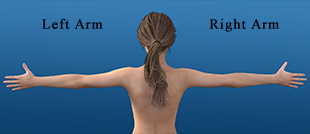
Where Does it Hurt?®
Not sure what service you need or what injury or syndrome you may have? Use our free, interactive tool to help you understand more about what you are experiencing. Start by clicking on the image below.
Virtual Consultation
Located out of the area? Dr. Knight may be able to help you virtually with an online virtual consultation.

Real Patient Reviews

“I don’t often write reviews for Doctors offices..But this office is really exceptional in terms of service and my wrist is now great! Which is really the most important thing.”
Featured On